Page 158 - 《精细化工》2020年第8期
P. 158
·1656·                            精细化工   FINE CHEMICALS                                 第 37 卷

                                                               [4]   FALDINI C, TRAINA F, PERNA F, et al. Surgical treatment of aseptic
                                                                   forearm nonunion with plate and opposite bone graftstrut. Autograft
                                                                   or allograft?[J]. International Orthopaedics, 2015, 39(7): 1343-1349.
                                                               [5]   LANGER  R,  VACANTI  J  P.  Tissue  engineering[J].  Science,  1993,
                                                                   260(5110): 920-926.
                                                               [6]   HE C L (贺超良), TANG Z H (汤朝晖), TIAN H Y (田华雨), et a1.
                                                                   Progress in the development of biomedical polymer materials fabricated
                                                                   by 3-dimensional printing technology[J]. Acta Polymerica Sinica (高
                                                                   分子学报), 2013, 44(6): 722-732.
                                                               [7]   BILLIET T, VANDENHAUTE M, SCHELFHOUT J, et a1. A review
                                                                   of  trends  and  limitations  in  hydrogel-rapid  prototyping  for  tissue
                                                                   engineering[J]. Biomaterials, 2012, 33(26): 6020-6041.
                                                               [8]   PEI  X,  MA  L,  ZHANG  B  Q,  et al.  Creating  hierarchical
                                                                   porosityhydroxyapatite  scaffold  with  osteoinduction  by  three-
                                                                   dimensionalprinting  and  microwave  sintering[J].  Biofabrication,
                                                                   2017, 9(4): 1-24.
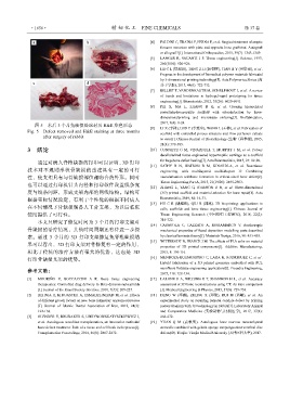
               图 5    术后 3 个月兔缺损处取材及 H&E 染色形态
            Fig. 5    Defect removed and H&E staining at three months   [9]   LI X (李祥), LI D C (李涤尘), WANG L (王林), et al. Fabrication of
                                                                   scaffold with controlled porous structure and flow perfusion culture
                   after surgery of rabbit
                                                                   in vitro[J]. Chinese Journal of Biotechnology (生物工程学报), 2005,
                                                                   21(4): 579-583.
            3   结论                                             [10]  CUNNIFFE  G  M,  VINARDELL  T,  MURPHY  J  M,  et al.  Porous
                                                                   decellularized tissue engineered hypertrophic cartilage as a scaffold
                 通过对病人脊椎缺损的打印可以证明,3D 打印                            for largebone defect healing[J]. Acta Biomaterialia, 2015, 23: 82-90.
                                                               [11]  SATHY  B  N,  WATSON  B  M,  KINARDLA,  et al.  Bonetissue
            技术对不规则形状骨缺损的重建具有一定的可行                                  engineering  with  multilayered  scaffolds-part  Ⅱ:  Combining
            性,使支架具有与骨缺损部位相吻合的外形。同时                                 vascularization  withbone  formation  in  critical-sized  bone  defect[J].
                                                                   Tissue Engineering Part A, 2015, 21(19/20): 2495-2503.
            也可以通过打印机针头内径和打印软件设置线条宽
                                                               [12]  ZHANG  L,  YANG  G,  JOHNSON  B  N,  et al.  Three-dimensional
            度与线条间距,形成支架内部的网线结构。结构可                                 (3D) printed scaffold and material selection for bone repair[J]. Acta
            根据实际情况设定,有利于个性化的根据不同病人                                 Biomaterialia, 2019, 84: 16-33.
                                                               [13]  HU  C  R  (胡超然), QIU B (邱冰).  3D  bioprinting:  applications  in
            在不同情况下骨缺损制备人工骨支架,为以后临床                                 cells,  scaffolds  and  bone  tissue  engineering[J].  Chinese  Journal  of
            使用提供了可行性。                                              Tissue  Engineering  Research  (中国组织工程研究),  2018,  22(2):
                 本文只研究了修复时间为 3 个月的打印支架对                            316-322.
                                                               [14]  CASAVOLA  C,  CAZZATO  A,  MORAMARCO  V.  Aorthotropie
            骨缺损的治疗情况,其他时间周期还有待进一步探                                 mechanical properties of fused deposition modelling parts described
            索。通过 3 个月的 3D 打印支架修复兔脊椎缺损结                             by classicallaminate theory[J]. Materials Design, 2016, 90: 453-458.
                                                               [15]  WITTBRODT B, PEARCE J M. The effects of PLA color on material
            果可以看出,3D 打印支架对骨修复有一定的作用,                               properties  of  3D  printed  components[J].  Additive  Manufacturing,
            相比于传统的治疗方法有很大的优势,这也是 3D                                2015, 8: 110-116.
            打印骨缺损支架的优势。                                        [16]  MENDOZA-BUENROSTRO C, LARA H, RODRIGUEZ C, et a1.
                                                                   Hybrid  fabrication  of  a  3D  printed  geometry  embedded  with  PCL
                                                                   nanofibers fortissue engineering applications[J]. Procedia Engineering,
            参考文献:
                                                                   2015, 110: 128-134.
            [1]   MOURIÑO  V,  BOCCACCINI  A  R.  Bone  tissue  engineering   [17]  LALONE E A, WILLING R T, SHANNON  H L,  et a1. Accuracy
                 therapeutics: Controlled drug delivery in three-dimensionalscaffolds   assessment of 3D bone reconstructions using CT: An intro comparison
                 [J]. Journal of the Royal Society Interface, 2010, 7(43): 209-227.   [J]. Medical Engineering & Physics, 2015, 37(8): 729-738.
            [2]   BEHNIA H, KHOJASTEH A, ESMAEELINEJAD M, et al. Effects   [18]  DENG  W  (邓威), ZHENG X (郑欣), RUI M (芮敏),  et al.  An
                 of different growth factors on new bone formation: asystem-aticreview   experimental study on repairing femoral condyle defect by printing
                 [J].  Journal  of  Islamic  Dental  Association  of  Iran,  2012,  24(3):   porous titanium with 3D technology in rabbits[J]. Laboratory Animal
                 122-138.                                          and  Comparative  Medicine  (实验动物与比较医学),  2017,  37(4):
            [3]   OLENDER E, BRUBAKER S, UHRYNOWSKATYSZKIEWICZ I,   266-272.
                 et al. Autologous osteoblast transplantation, an innovative methodof   [19]  YUAN  Q  M  (袁秋明).  Autologous  bone  marrow  mesenchymal
                 bone defect treatment: Role of a tissue and cell bank in theproces[J].   stemcells combined with gelatin sponge torepairgoatintervertebral disc
                 Transplantation Proceedings, 2014, 46(8): 2867-2872.   defects[D]. Tianjin: Tianjin Medical University (天津医科大学), 2017.

